
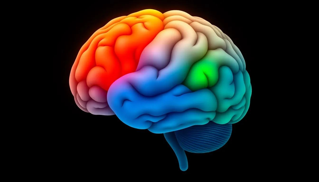
When people talk about trauma “changing” someone, they’re more right than they realize. Trauma doesn’t just change how you feel — it physically changes how your brain functions. The fear circuits become hyperactive. The rational thinking centers go quiet. The memory systems misfire, replaying the past as though it’s happening right now.
Understanding these changes isn’t just academically interesting — it’s clinically important. When you know what trauma does to the brain, PTSD symptoms stop seeming like personal weakness and start making neurological sense. And most importantly, these changes are reversible. The same brain plasticity that allows trauma to reshape neural circuits also allows treatment to restore them.
PTSD prevalence in the United States affects approximately 6% of the population at some point in their lifetime. Behind that number are millions of brains that have been reshaped by traumatic experience — and that can be reshaped again through evidence-based treatment.
The Brain’s Alarm System: How the Threat Response Works
To understand what trauma changes, you first need to understand how the brain handles threats under normal conditions.
When you encounter danger — a car swerving into your lane, a loud explosion, a physical attack — your brain activates a rapid-response system designed to keep you alive. This happens in milliseconds, far faster than conscious thought:
- Sensory input arrives at the thalamus (the brain’s relay station)
- The amygdala receives the signal and makes an instant assessment: Is this dangerous?
- If yes, the amygdala triggers the fight-or-flight response — flooding the body with adrenaline and cortisol
- The hypothalamic-pituitary-adrenal (HPA) axis activates, sustaining the stress response
- Heart rate spikes, muscles tense, breathing quickens, pain sensitivity drops
- The prefrontal cortex — the rational, thinking brain — evaluates the situation more slowly and can modulate the response: Was that actually a threat, or just a car backfiring?
- Once safety is confirmed, the prefrontal cortex tells the amygdala to stand down, cortisol levels normalize, and the body returns to baseline
- The hippocampus files the memory with context: This happened at 3 p.m. on Oak Street. It’s over now. I’m safe.
This system works brilliantly for discrete threats. The problem arises when the threat is overwhelming, prolonged, or repeated — and the system can’t complete its cycle.
Three Brain Regions Changed by Trauma
Decades of neuroimaging research have identified a consistent pattern of brain changes in people with PTSD. A landmark review by Bremner (2006), cited over 1,300 times, identified alterations in a circuit involving three key structures: the medial prefrontal cortex, the hippocampus, and the amygdala (Bremner, 2006).
These aren’t subtle differences. They’re measurable, reproducible changes in both brain structure (volume) and function (activity levels) that directly correspond to the symptoms people with PTSD experience.
The Amygdala: A Hyperactive Alarm
What it does normally: The amygdala is the brain’s threat detector. It processes fear, detects danger, and initiates the stress response. It’s fast, automatic, and operates below conscious awareness.
What trauma does to it: In PTSD, the amygdala becomes hyperactive — stuck in a state of heightened reactivity. It fires more easily, more intensely, and in response to stimuli that aren’t actually dangerous.
Brain imaging studies consistently show that people with PTSD exhibit significantly greater amygdala activation in response to trauma-related cues compared to people without PTSD. But the hyperactivation isn’t limited to trauma reminders — the amygdala also overreacts to neutral stimuli, general emotional content, and even ambiguous situations (Bremner, 2006).
How this creates symptoms:
- Hypervigilance: The alarm system is always scanning for threats, keeping you in a state of constant alertness
- Exaggerated startle response: A door slamming, a car horn, someone tapping your shoulder — the amygdala reacts as if it’s a mortar round
- Emotional reactivity: Small frustrations trigger disproportionate anger, fear, or panic
- Sleep disruption: The amygdala doesn’t fully deactivate during sleep, producing nightmares and fragmented rest
- Avoidance: The amygdala associates certain people, places, sounds, and smells with danger, driving powerful avoidance behavior
The amygdala isn’t malfunctioning — it’s doing exactly what it was designed to do. The problem is that it was calibrated for a war zone and hasn’t recalibrated for peacetime.
The Prefrontal Cortex: Losing the Brake Pedal
What it does normally: The prefrontal cortex (PFC) — particularly the medial PFC, including the anterior cingulate cortex — is responsible for executive function: rational thinking, emotional regulation, decision-making, and impulse control. Critically, it’s the brain region that can tell the amygdala to calm down.
What trauma does to it: In PTSD, the prefrontal cortex becomes hypoactive — it’s underperforming precisely when it’s needed most. Research by Kredlow et al. (2022), published in Neuropsychopharmacology and cited over 400 times, confirmed that the prefrontal cortex is “central to fear processing” and that its regulation of the amygdala is impaired in PTSD (Kredlow et al., 2022).
UAB Medicine describes this dynamic clearly: “The prefrontal cortex regulates emotions and decision-making but can be impaired in PTSD, making it harder to control fear” (UAB Medicine, 2024).
How this creates symptoms:
- Inability to rationalize safety: You know intellectually that you’re safe, but you can’t make yourself feel safe — the prefrontal cortex can’t override the amygdala’s alarm
- Emotional dysregulation: Without the PFC’s moderating influence, emotions swing between extremes — rage, terror, numbness — with little middle ground
- Difficulty concentrating: The PFC governs attention and working memory, both of which suffer when it’s suppressed
- Impaired decision-making: The executive functions needed for planning, prioritizing, and problem-solving are diminished
- Reduced self-awareness: The PFC helps you observe your own mental states. When it’s underactive, you may not recognize that you’re spiraling until you’re deep in a flashback or rage
Think of it this way: the amygdala is the gas pedal, and the prefrontal cortex is the brake. In PTSD, someone is flooring the gas while the brake lines have been cut.
The Hippocampus: When Memory Misfires
What it does normally: The hippocampus is essential for forming, organizing, and retrieving memories. It gives memories context — time stamps, locations, the sequence of events. It’s what allows you to remember something as past rather than experiencing it as present.
What trauma does to it: The hippocampus is particularly vulnerable to chronic stress. Elevated cortisol levels — sustained during prolonged or repeated trauma — are toxic to hippocampal neurons. Neuroimaging studies consistently find reduced hippocampal volume in people with PTSD (Bremner, 2006). Research from NHS Wales confirms that “adults with PTSD have a reduction in the volume of the hippocampus” (NHS Wales, 2024).
How this creates symptoms:
- Flashbacks: Without proper hippocampal processing, traumatic memories aren’t stored with context. When triggered, the memory replays as a raw sensory experience — sights, sounds, smells, bodily sensations — without the hippocampal “time stamp” that says this is a memory from 2019, it’s not happening now
- Fragmented memory: Traumatic memories are often stored in pieces — vivid sensory fragments without a coherent narrative. You might remember the smell of burning rubber and the sound of screaming but not the sequence of events
- Difficulty forming new memories: Chronic cortisol exposure impairs the hippocampus’s ability to consolidate new information, contributing to the “brain fog” many people with PTSD describe
- Difficulty distinguishing past from present: Without proper contextual encoding, the brain treats past trauma as an ongoing threat — the alarm never fully resolves
How These Changes Create PTSD Symptoms
When you map brain changes to symptoms, the entire PTSD presentation makes neurological sense:
| Symptom | Brain Region | Mechanism |
|---|---|---|
| Flashbacks | Hippocampus | Memories lack time-context, replay as present |
| Hypervigilance | Amygdala | Threat detection permanently elevated |
| Emotional numbness | Prefrontal cortex | Shutdown as protective mechanism |
| Nightmares | Amygdala + hippocampus | Fear circuits active during sleep, memory fragments replay |
| Avoidance | Amygdala | Conditioned fear response to trauma-associated cues |
| Irritability/rage | Amygdala + PFC | Hyperactive alarm + impaired emotional braking |
| Concentration problems | PFC + hippocampus | Executive function suppressed, memory encoding impaired |
| Exaggerated startle | Amygdala | Lowered threshold for threat detection |
This isn’t a character flaw. It’s a brain injury caused by experience. Veterans experience these neurological changes at higher rates due to prolonged combat exposure, and children exposed to trauma may experience more severe changes because their brains are still developing.
Neuroplasticity: The Brain Can Heal
The most important fact about trauma and the brain is this: the same plasticity that allows trauma to change the brain also allows treatment to change it back.
Neuroplasticity — the brain’s ability to reorganize itself by forming new neural connections — doesn’t stop after trauma. It continues throughout life. And evidence-based PTSD treatments leverage this plasticity directly:
How CPT works neurologically: Cognitive Processing Therapy targets the prefrontal cortex. By helping patients identify and challenge distorted beliefs about the trauma, CPT strengthens prefrontal activity and its ability to regulate the amygdala. Over sessions, the brake pedal gets reconnected.
How EMDR works neurologically: EMDR appears to facilitate hippocampal processing of traumatic memories. The bilateral stimulation during memory recall may help the hippocampus properly contextualize and file the traumatic memory — adding the time stamp that says “this happened then, not now.”
How Prolonged Exposure works neurologically: PE directly targets amygdala hyperactivation through extinction learning. By repeatedly confronting trauma-related stimuli in a safe environment, the amygdala gradually learns that these cues no longer signal danger. The alarm recalibrates.
The evidence for neural recovery:
- Studies show that successful PTSD treatment is associated with increased prefrontal cortex activity and decreased amygdala hyperactivation
- Hippocampal volume can partially recover with treatment and stress reduction
- These neural changes correlate with symptom improvement — patients who show greater prefrontal changes tend to show greater clinical improvement
For a detailed guide to these treatments, see our article on PTSD treatment options that target these brain changes.
What you can do alongside treatment:
- Exercise: Aerobic exercise promotes neurogenesis (new neuron growth) in the hippocampus and increases BDNF (brain-derived neurotrophic factor)
- Sleep: Restorative sleep is essential for memory consolidation and neural repair
- Mindfulness meditation: Regular practice has been shown to reduce amygdala reactivity and increase prefrontal cortex thickness
- Social connection: Safe, supportive relationships help regulate the stress response system
The brain changed because of what happened to you. It can change again because of what you do next.
Frequently Asked Questions
Can trauma permanently damage the brain?
Trauma causes measurable changes in brain structure and function, but “permanent damage” is misleading. While chronic, untreated PTSD can lead to persistent changes — including reduced hippocampal volume and chronic amygdala hyperactivation — these changes are at least partially reversible with treatment. Neuroplasticity allows the brain to form new connections and patterns throughout life. Studies show that successful PTSD treatment is associated with normalization of brain activity in affected regions.
What part of the brain is most affected by PTSD?
Three regions are most consistently affected: the amygdala (becomes hyperactive, driving fear and hypervigilance), the prefrontal cortex (becomes hypoactive, impairing emotional regulation and rational thinking), and the hippocampus (volume decreases, causing memory fragmentation and flashbacks). These three structures form a circuit, and the disruption of their normal interaction produces the core PTSD symptom clusters.
Can you see PTSD on a brain scan?
Research brain imaging can detect group-level differences between people with and without PTSD — patterns of amygdala hyperactivation, prefrontal hypoactivation, and reduced hippocampal volume. However, brain scans are not currently used for clinical PTSD diagnosis. The variation between individuals is too great for a scan to reliably diagnose one person. PTSD remains a clinical diagnosis based on symptoms, history, and functional impact.
How does childhood trauma affect brain development?
Childhood trauma can have particularly severe effects because the brain is still developing. The prefrontal cortex doesn’t fully mature until the mid-20s, so trauma during childhood or adolescence can disrupt its development, potentially leading to more pronounced difficulties with emotional regulation, impulse control, and executive function. Childhood trauma also affects the developing stress response system, potentially calibrating it for a threatening world — which increases vulnerability to PTSD, depression, and anxiety later in life.
Does the brain heal on its own after trauma, or is treatment necessary?
Some people do recover naturally after traumatic events — the brain’s stress response system resolves on its own within weeks to months. But for those who develop PTSD (approximately 6% of the population after trauma exposure), spontaneous recovery is uncommon without intervention. The average untreated PTSD episode persists for years. Evidence-based treatments like CPT, EMDR, and Prolonged Exposure actively leverage neuroplasticity to restore healthy brain function — they don’t just manage symptoms, they address the underlying neural changes.
Sources
-
Bremner JD. Traumatic stress: effects on the brain. Dialogues in Clinical Neuroscience. 2006;8(4):445-461. https://pmc.ncbi.nlm.nih.gov/articles/PMC3181836/
-
Kredlow MA, et al. Prefrontal cortex, amygdala, and threat processing: implications for PTSD. Neuropsychopharmacology. 2022;47:247-259. https://www.nature.com/articles/s41386-021-01155-7
-
UAB Medicine. Inside the mind: what PTSD does to the brain. University of Alabama at Birmingham; 2024. https://www.uab.edu/medicine/news/psychiatry/inside-the-mind-what-ptsd-does-to-the-brain
-
VA National Center for PTSD. PTSD treatment basics. U.S. Department of Veterans Affairs; Updated September 2025. https://www.ptsd.va.gov/understand_tx/tx_basics.asp
-
American Psychological Association. Treatments for PTSD — clinical practice guideline. APA; 2025. https://www.apa.org/ptsd-guideline/treatments
-
National Institute of Mental Health. Depression. NIMH; Revised 2024. https://www.nimh.nih.gov/health/publications/depression
-
World Health Organization. Mental disorders — fact sheet. WHO; 2024. https://www.who.int/news-room/fact-sheets/detail/mental-disorders
-
Traumatic Stress Wales. Trauma and the brain. NHS Wales; 2024. https://traumaticstress.nhs.wales/children-and-young-people/trauma-and-the-brain/